Supporting orthopedic evaluation with AI-assisted bone angle calculation.
X-ray to 3D CT Reconstruction & Knee Alignment Analytics
Traditional CT scans are several times more expensive than X-rays, often delaying critical orthopedic diagnosis. Our client needed a system to reconstruct 3D CT-grade results from standard 2D X-rays to calculate precise bone alignment angles for surgical planning.
Client
Hospital based in the US
Year
2024
Location
USA
Domain
Computer Vision
The Challenge
Orthopedic evaluation for lower limb alignment typically requires expensive 3D imaging to achieve necessary precision. Converting a 2D X-ray into a volumetrically accurate 3D CT scan—while maintaining clinical-grade sub-millimeter precision—is a monumental technical challenge.
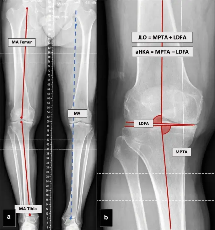
The system needed to reliably identify and measure bone angles such as the Lateral Distal Femoral Angle (LDFA) and Medial Proximal Tibial Angle (MPTA) as a tool for initial screening. Generalizing this across diverse patient anatomy with approximately 75-80% accuracy required a specialized deep learning approach that prioritizes diagnostic consistency.
2D to 3D Spatial Mapping
Translating flat X-ray perspectives into volumetrically accurate 3D models.
Reconstruction Precision
Developing a model that produces reliable 3D estimates (75-80% accuracy) from 2D data.
Anatomical Variability
Ensuring the model generalizes across diverse patient knee structures and conditions.
Cost-Efficiency vs Quality
Delivering CT-grade diagnostic value using low-cost X-ray imaging.
Our Solution
We engineered a deep learning pipeline centered on 2D to 3D image registration and generative reconstruction. By training on paired medical datasets, the system learned to predict three-dimensional osseous geometry from flat projections, followed by a dedicated analytics layer for automated orthopedic measurement.
GAN-Powered Reconstruction
We developed and trained a neural network model using Generative Adversarial Networks (GANs) to reconstruct high-fidelity 3D bone structures from 2D X-ray inputs.
- Anti-hallucination training
- Anatomical integrity focus
- High-resolution output
Automated Angle Analytics
We built an engine to automatically calculate critical parameters like CPAK, mHKAA, LDFA, and MPTA for orthopedic evaluation.
- Precise alignment discovery
- Standardized measurements
- Overlaid visual results
Anatomic Feature Detection
The system identifies key landmarks including femoral condyles and the functional flexion-extension (F/E) axis with extreme accuracy.
- Automated bone segmentation
- Landmark pinpointing
- Surgical axis identification
Cloud-Native Infrastructure
To ensure fast clinical turnaround, we deployed the entire deep learning pipeline on high-performance AWS instances.
- Near real-time processing
- Scalable data ingestion
- Secure DICOM-ready hosting
The Impact
Providing 3D diagnostic assistance for initial screenings at the cost of 2D imaging has effectively lowered the barrier for early orthopedic evaluation and preoperative planning.
CT-level insights at X-ray costs
Automated measurement precision
Faster preoperative planning
Project Showcase
Observe the AI assisted bone angle calculation in action, supporting initial diagnosis through automated orthopedic evaluation.
Dynamic Angle Calculation Engine
Similar Case Studies
Start Your AI Consulting Services Partnership
Ready to take the first step towards unlocking opportunities, realizing goals, and embracing innovation? We're here and eager to connect.
Phone
+1 (737) 381-3933
Phone
+92 (314) 424-5425
USA
5900 Balcones Drive, STE 100 Austin TX 78731
Pakistan
585-H3, Johar Town, Lahore (Opp. Expo Center)